CRISPR-Cas9, cutting DNA
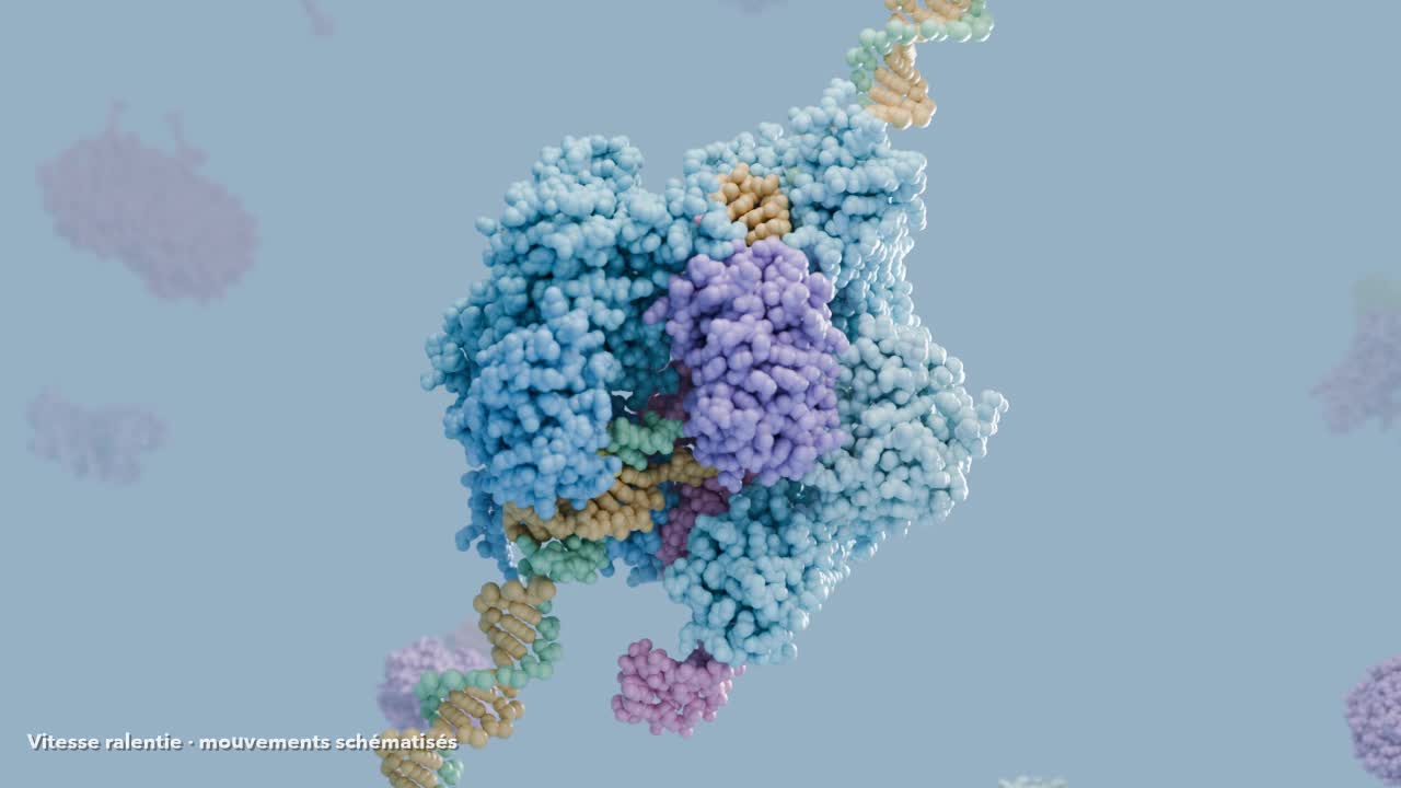
The 3D animation from the video, replayable and hands-on: Cas9 looks for the PAM, opens the DNA, and cuts. Pause, hide parts, move them, then put everything back.
Watch the video · The next CRISPR?Variables
- Playback: the animation from the video, at normal speed, slowed down or sped up, step by step
- The parts: each Cas9 domain, the guide RNA and both DNA strands, to show or hide
- The position of the parts: move them, spread them apart, then put them back
- The camera: the video’s own, or free (rotate, zoom)
Key figures
- 20 nt
- length of the guide RNA spacer
- NGG
- PAM recognised by SpCas9
- 3 bp
- distance between the PAM and the cut
- 1,368
- amino acids in SpCas9 (≈ 160 kDa)
- 4
- real structures from the Protein Data Bank
What happens, step by step.
- 01
An RNA that acts as a guide
Cas9 is a protein from the bacterium Streptococcus pyogenes. On its own, it does not know where to cut. It carries a guide RNA (in the bacterium it is made of two RNAs; researchers fused them into one) whose first 20 letters, the spacer, are chosen by the researcher: they reproduce the DNA sequence to target.
- 02
The PAM, a pass
Cas9 does not read the whole genome letter by letter. It first looks for a short motif, the PAM (NGG for S. pyogenes Cas9), with its PI domain. Without a PAM right next to it, even a perfectly complementary sequence is ignored. This is also what protects the bacterium: in its own CRISPR array, spacers are not followed by a PAM.
- 03
The R-loop
Once the PAM is recognised, Cas9 separates the two strands right next to it. The guide RNA pairs with the target strand, letter by letter, starting from the PAM. The non-target strand is pushed away: this is the R-loop. The first 10 to 12 pairs, the seed region, must be perfect; a mismatch there stops the opening.
- 04
Two blades, one break
When pairing is complete, the HNH domain swings over and cuts the target strand; the RuvC domain cuts the non-target strand. Both cuts fall 3 base pairs before the PAM: the DNA is cut cleanly, straight across.
- 05
The cell repairs, and that is where editing happens
Cas9 changes nothing itself: it breaks. The cell rejoins the ends. Direct joining (NHEJ) is often exact, but Cas9 cuts again as long as the target is intact: eventually a few letters are added or lost, which can knock out the gene. If a DNA template is supplied, repair by recombination (HDR) can copy in a chosen sequence.
What the scene simplifies
- This is the animation from the video: speeds are changed and movements are schematic.
- Cas9 domains are rigid bodies, moved between the poses of four real structures (4ZT0, 4UN3, 5F9R, 6O0Y): a morph, not a simulation.
- DNA is shown as beads (phosphate, sugar, base), fitted to the 5F9R crystal and extended with an ideal helix; the part of the non-target strand not resolved in the crystal is a schematic arc.
- The PAM search is summed up in two stops; in reality Cas9 diffuses in 3D and probes a great many PAMs.
- Each atom is a van der Waals sphere; hydrogens are not shown. Colours are readability conventions.
- The background proteins are real structures, placed to evoke the crowding of the cell.
Further reading
- Jinek et al., Science 337, 816 (2012)
Cas9 programmed by a guide RNA; cut 3 bp upstream of the PAM.
- Nishimasu et al., Cell 156, 935 (2014)
Crystal structure of SpCas9; REC and NUC lobes, HNH, RuvC and PI domains.
- Sternberg et al., Nature 507, 62 (2014)
Target search by PAM interrogation; seed region.
- Jiang et al., Science 348, 1477 (2015)
Cas9 loaded with its guide RNA (PDB 4ZT0).
- Zhu et al., Nat. Struct. Mol. Biol. 26, 679 (2019)
HNH domain swinging toward the target strand, with magnesium (PDB 6O0Y).
- Anders et al., Nature 513, 569 (2014)
PAM recognition by the PI domain (PDB 4UN3).
- Jiang et al., Science 351, 867 (2016)
Structure of the R-loop and HNH domain movement (PDB 5F9R).
Can an AI reinvent genetic scissors?